13 Haziran 2014 Cuma

NEUROCHEMISTRY

Source: http://www.humanneurophysiology.com/neurochemistry.htm

Electron microscope studies have shown that capillaries in the brain have a continuous capillary endothelium with tight junctions and are therefore unlike the more permeable capillaries found elsewhere in the body. This effectively excludes the passage of many materials including proteins and molecules with molecular weights as low as 2000. The existence of the barrier was first demonstrated by Paul Ehrlich and later by Goldman, who in 1909 injected large amounts of the dye trypan blue into the vascular circulation and observed that all tissues became intensely stained while the brain remained "snow white." We now know that trypan blue was excluded from the brain because it rapidly complexed with albumen in the plasma and could not cross cerebral capillaries in this form. Many materials do, however, cross the barrier from plasma to brain. Nevertheless, it's a "selective" barrier where some materials are excluded or cross with difficulty and others pass quite freely. Because of the heavy dependence of the brain on a steady supply of oxygen for cellular respiration and glucose for energy metabolism, it is not surprising to find that they pass freely into the brain with little hindrance. Similarly, metabolic wastes and carbon dioxide readily pass across the barrier from brain to plasma. On the other hand, free fatty acids, an easily accessible alternate energy source for most other cells of the body including muscle, are virtually excluded from the brain. A summary of the permeability of the blood-brain barrier to several different metabolic substances is illustrated in Fig. -1
bulletDiffusion Across the Barrier

O2, CO2, N2O, Kr, and Xe are gases which readily diffuse across the barrier. The latter three have been used to calculate cerebral blood flow. Water also readily diffuses into and out of the brain. Its net movement is dictated solely by the osmolality of the plasma. Thus an increase in the plasma osmolality from its normal value of 290 mosmol/L can draw water from the brain by osmosis, and actually shrink its volume. This phenomenon has been employed clinically to reduce intracranial pressure by using plasma expanders such as mannitol to increase plasma osmolality. Mannitol does not cross the blood-brain barrier.
Lipid solubility is an important factor in diffusion across the barrier. Generally the higher the lipid solubility of a substance, the more readily it diffuses. Thus alcohols like ethanol move freely into the brain. Lipid-soluble thiobarbital equilibrates more rapidly between plasma and brain than the slightly less soluble barbital does. Salicylic acid is less soluble yet and thus requires even more time to equilibrate. 
bulletFacilitated Transport across the Barrier
Carrier systems appear to be involved in the transport of several materials across the barrier. Glucose, ions, and certain amino acids utilize this type of system. The carrier system for glucose is stereospecific as n-glucose, but not L­glucose, is readily transported into the brain. Lactic, pyruvic, and acetic acids also utilize such carriers.
While proteins are virtually excluded from the brain, certain amino acids  pass readily into it. Included are the essential amino acids and those which are precursors for the production of neurotransmitters. The latter include tyrosine (required for norepinephrine and dopamine synthesis) and tryptophan (for serotonin synthesis). Similarly, neuroactive peptides whose amino acid sequences have been clearly identified such as substance P, methionine enkephalin and leucine enkephalin, ,B-endorphin, ACTH, angiotensin II, oxytocin, vasopressin, somatostatin, thyrotropin-releasing factor, and luteinizing hormone-releasing factor rely on a steady transport of these amino acids from plasma to brain for their continued synthesis.
Ions cross the barrier into brain but do so much more slowly than into other body tissues. An intravenous K + administration exchanges much more quickly with muscle tissue that it does with brain. Ca2+ and Mg2+ transport is equally slow, while N a + is somewhat faster. Hion transport into the brain is very slow.
Certain areas of the brain apparently contain no blood-brain barrier. These include the neurohypophysis, median eminence of the hypothalamus, the area postrema, and the pineal gland. Because many circulating hormones control their own release through negative feedback to the hypothalamus, the importance of barrier lack in this area is readily apparent. If such hormones are to influence the hypothalamic output of releasing or inhibiting factors to the anterior pituitary via the hypothalamohypophyseal portal system, they must not be barred from the hypothalamus by a barrier system. Similarly, osmoreceptors of the hypothalamus must be able to constantly and easily detect changes in the osmolality of the plasma if the release of antidiuretic hormone (ADH) is to proceed properly.


bulletCEREBRAL BLOOD FLOW
The average cerebral blood flow in humans is approximately 55 mL per 100 g of brain tissue per minute. This is a little over 700 mL/min for a 1350-g brain. Thus while the human brain comprises only about 2.5 percent of the body's weight, it receives almost 15 percent of the cardiac output, attesting to the high vascular demands of this organ.
A reliable and frequently used method of determining cerebral blood flow is the method of Kety and Schmidt. It is based on the Fick principle and utilizes the arteriovenous difference of a freely diffusible gas such as N2O as it passes through the brain. Accordingly, the flow of blood through the brain can be determined by measuring the amount of N2O removed from the blood by the brain per minute and dividing this by the arteriovenous difference of N2O as it passes through the brain. The cerebral blood flow is higher in children than in adults, typically exceeding 100 mL per 100 g per minute. However, contrary to popular thinking, the blood flow decreases only slightly with advancing age. The brain utilizes fully 25 percent of the body's total oxygen consumption. The arteriovenous O2 difference is relatively high since the brain receives only 15 percent of the cardiac output. The arteriovenous difference is 6.6 mL per 100 ml., falling from 19.6 to 13 mL per 100 mL as blood passes through the brain (Fig. 17-2). Thus we can calculate a cerebral oxygen consumption of approximately 3.5 mL per 100 g per minute. This value is greater in skeletal muscle, skin. and liver. but less in cardiac muscle and kidney.

bullet
OXYGEN CONSUMPTION
The utilization of oxygen by the brain is not uniform throughout its mass. The gray matter consumes as much as 94 percent of cerebral oxygen. while the white matter, which makes up fully 60 percent of the brain's mass. consumes only 6 percent. Oxygen consumption, and hence oxygen need, increases as we move up the neuraxis. It is lowest in the spinal cord and increases through the medulla. midbrain, thalamus, cerebellum, and cerebral cortex. Thus it is not surprising to find that the sensorimotor functions of the cerebral cortex are more sensitive to hypoxic damage than are the vegetative functions of the pontomedullary areas. Progressive decreases in cerebral oxygen consumption are always accompanied by progressive decreases in the level of mental alertness. Compared to the mentally alert young man with an O2 consumption of 3.5 mL per 100 g per minute, the mentally confused states associated with diabetic acidosis, insulin hypoglycemia, and some forms of cerebral arteriosclerosis might typically show O2 consumptions rates down to 2.8 mL per 100 g per minute. Finally, the comatose states of diabetic coma. insulin coma. and anesthesia can show consumption rates as low as 2.0 mL per 100 g per minute. On the other hand. O2 consumption by the brain increases during convulsions.

bulletEFFECTS OF OXYGEN DEPRIVATION
Almost all of the oxygen consumed by the brain is utilized for the oxidation of carbohydrate. Sufficient energy is released from this process so that the normal level of oxygen utilization is adequate to replace the 12 mmol or so of A TP which the whole brain uses per minute. However, since the normal brain reserve of A TP and creatine phosphate (CrP) totals only about 8 rnmol, less than a minute's reserve of high energy phosphate bonds is actually available if production were to suddenly stop. In the absence of oxygen, the anerobic glycolysis of glucose and glycogen could supply only another 15 mmol of A TP, as these two energy substrates are stored in such low quantities in brain tissue.
A continuous uninterrupted supply of oxygen to the brain is essential in order to maintain its metabolic functions and to prevent tissue damage. The ox­ygen-independent glycolytic pathway (anerobic glycolysis) is insufficient, even at maximum operating levels, to supply the heavy demands of the brain. Thus a loss of consciousness occurs when brain tissue P02 levels fall to 15 to 20 mmHg. This level is reached in less than 10 s when cerebral blood flow is com­pletely stopped
Low tissue oxygen levels in the brain (hypoxidosis) can be caused by de­creased blood flow (ischemia) or with adequate blood flow accompanied by low levels of blood oxygen (hypoxemia). It is important to recognize that decreased P02 caused by ischemia is accompanied by decreased brain glucose and in­creased brain CO2 while hypoxemia with normal blood flow is not accompanied by changes in brain glucose or CO2with complete cessation of CBF, irreversible damage occurs to brain tissue within a few minutes and the histological ef­fects observed are remarkably similar whether caused by ischemia, hypoxemia, or hypoglycemia.
Experimental studies on rats and mice in which arterial P02 is progres­sively reduced have illustrated some aspects of hypoxemia which are likely to be similar in humans. A drop in arterial P02 to 50 mmHg (normal, 96 mmHg) produces no change in CBF, O2 utilization by the brain, or lactic acid produc­tion. However, as P02 levels drop to 30 mmHg, a 50 percent increase in CBF is observed along with the onset of coma, decreased oxygen utilization, and in­creased lactic acid production. When the P02 drops further to 15 mmHg, 50 percent of the animals die because of cardiac failure. The remainder show a tremendous increase in lactic acid production, but, surprisingly, levels of ATP, ADP, and AMP remain normal. If cerebral perfusion is artificially maintained while the arterial P02 is decreased further, ATP, ADP, and AMP levels still remain normal. The implication is that the coma observed at low oxygen levels may not be due to a decrease in ATP but instead to some still unexplained mechanism. It appears likely that cardiac complications caused by hypoxemia and the subsequent effect on cerebral blood flow may actually be a primary cause of the irreversible pathologic damage to the brain.
Hypoxia, such as that brought on by high altitudes, brings on a number of symptoms, including drowsiness, apathy, and decreases in judgment. Unless oxygen is administered within half a minute or so, coma, convulsions, and depression of the EEG occur.


 Glucose metabolism

Glucose is virtually the only energy substrate which the brain can use. Free fatty acids, used by most other tissues when glucose is in short supply, are excluded from the brain by the blood-brain barrier. The brain extracts 6.6 mL of O2 from each 100 mL of cerebral blood and returns 6.7 mL of CO2. Thus the respiratory quotient (RQ) of the brain is approximately 1.0, indicating carbohydrate utilization only. Brain glucose consumption is normally about 10 mg per 100 ml., accounting for almost 75 percent of the liver's production and further attesting to the brain's heavy dependence on glucose. Adenosine triphosphate (ATP), produced by the metabolic degradation and oxidative phosphorylation of glucose, is the useful energy currency in brain tissue. About 85 percent of the circulating glucose extracted from the cerebral arterial blood is converted to CO2 via the tricarboxylic acid (TCA) cycle, while 15 percent is converted to lactic acid. The general scheme for glucose metabolism in the brain is similar to that in other tissues and is illustrated in Fig.1. The enormous ATP requirements of the brain are partly due to neurotransmitter synthesis, release, and reuptake as well as intracellular transport and complex synthetic mechanisms. But undoubtedly the greatest percentage of ATP is utilized to power the ion pumps which restore membrane potentials, enabling neurons to maintain their excitability.
     
Fig-1

 EFFECTS OF GLUCOSE DEPRIVATION
In the healthy normal functioning brain, glucose is the only substrate utilized for energy metabolism. Thus hypoglycemia presents the brain with a very serious problem. While most other tissues can shift to utilizing free fatty acids (FFA) as an alternative energy source when glucose is lacking, the brain cannot because they are excluded by the blood-brain barrier. While there is some evidence that the brain can utilize β-hydroxybutyric acid for energy metabolism when glucose levels are low or when fats are being mobilized for energy metabolism throughout the rest of the body, the brain could never supply its high energy demands by this method alone in the absence of glucose. Thus the brain is dependent on an uninterrupted supply of blood-borne glucose to energize its cells.
Decreases in blood glucose bring on disturbances in cerebral function. Depending on the level of hypoglycemia, these changes range from mild sensory disturbances to coma. At blood glucose levels of 19 mg per 100 mL or below (normal is 60 to 120 mg per 100 mL), a mentally confused state occurs. Brain O2 utilization falls to 2.6 mL per 100 g per minute (normal, 3.5 mL per 100 g per minute) and glucose utilization drops as well. Coma commences when glucose levels fall to 8 mg per 100 mL.
Epinephrine can be effective in reversing the effects of hypoglycemia by promoting liver glycogenolysis. However, attempts to solve the problem by substituting other carbohydrate metabolic substrates have been largely unsuccessful, with the single exception of mannose. This is the only monosaccharide other than glucose which the brain appears to utilize directly. It crosses the blood-brain barrier and directly replaces glucose in the glycolytic pathway. However, its normal level in the blood is too low to be of any real help in reversing the cerebral effects of hypoglycemia. Unless reversed quickly, comatose levels of prolonged hypoglycemia will bring on necrosis of cerebrocortical cells and (to a lesser extent) other brain regions as well.


Neurons in the CNS produce a large number of special molecules which function as neurotransmitters or are suspected to do so, including acetylcholine (ACh), norepinephrine (NE), dopamine (DA), y-aminobutyric acid (GABA), aspartic acid, glutamic acid, glycine, and substance P. CNS neurons also syn­thesize a number of neuropeptides which perform quite specific endocrine roles. We will take a closer look at these neuroactive chemicals now.
 Acetylcholine
 Acetylcholine has long been recognized as an important neurotransmitter. It's released by preganglionic sympathetic and parasympathetic nerve fiber termi­nals as well as postganglionic parasympathetic and certain select sympathetic fibers. It is also the only recognized neurotransmitter at the skeletal muscle neuromuscular junction. Unfortunately we don't have nearly as complete a pic­ture of the distribution of cholinergic neurons in the CNS. There appear to be cholinergic fibers associated with the arousal or activating systems of the brain which project from the midbrain reticular formation, hypothalamus striatum, and septum to the neocortex. ACh and the enzymes necessary for its synthesis are also found in the hippocampus, corpus striatum, and retina.
 Acetylcholine is formed by the reaction of choline with acetyl coenzyme A (acetyl CoA) in the presence of the enzyme choline acetyltransferase (CAT). Since neurons can't synthesize choline, the ultimate source of choline for ACh synthesis is the choline of the plasma. Acetyl CoA is synthesized within presynaptic cytoplasm by the A TP-energized reaction of acetate with CoA. Once ACH has been synaptically released and has produced its postsynaptic effects on membrane permeability, it is hydrolyzed within microseconds by the enzyme acetylcholinesterase (AChE). Interestingly enough, while negligible amounts of ACh are reabsorbed by presynaptic terminals in the peripheral ner­vous system (hydrolysis by AChE being overwhelmingly dominant), its reuptake by the terminals in brain is considerable. Nevertheless, its failure to be resequestered into synaptic vesicles leaves the significance of this process in doubt.
Acetate + CoA + ATP ~ acetyl CoA + AMP + 2Pi
 Catecholamines
 The catecholamine neurotransmitters are norepinephrine (NE) and dopamine (DA). The synthesis of both of these amines proceeds from the amino acid tyrosine (Fig. 17-5). Tyrosine is converted to 3,4-dihydroxyphenylalanine (dopa) by the enzyme tyrosine hydroxylase. Subsequent decarboxylation by dopa decarboxylase converts dopa to 3,4-dihydroxyphenylethylamine (dopa­mine). This is as far as the synthesis proceeds in dopaminergic neurons. In norepinephrinergic neurons, an additional step converts dopamine to norepi­nephrine by action of the enzyme dopamine ,B-hydroxylase.
 The enzymatic degradation of the two catecholamines is illustrated in Fig. 17-6. Catechol-o-methyltransferase (COMT) and monoamine oxidase (MAO) produce inactive products which have little effect on receptor sites. MAO catalyzes the oxidative deamination of norepinephrine to 3,4-dihydrox­ymandelic acid and dopamine to 3,4-dihydroxyphenylacetic acid. These prod­ucts are then methylated by COMT to 3-methoxy-4-hydroxymandelic acid and homovanillic acid, respectively. Alternatively, norepinephrine can first be methylated to normetanephrine and then deaminated to 3-methoxy-4-hydroxymandelic acid.
 Distribution of Norepinephrinergic Fibers
The distribution of norepi­nephrinergic fibers in the peripheral nervous system is limited to the majority of postganglionic sympathetic neurons. Norepinephrine-releasing neurons in the central nervous system have their cell bodies located in the midbrain, pons, and medulla, primarily in the reticular formation. Two norepinephrine systems are often described in the mammalian brain: the locus ceruleus system and the lateral tegmental system. The cell bodies of the former are located in the locus ceruleus, a prominent nucleus in the brain stem reticular formation at the level of the isthmus. This nucleus is composed entirely of norepinephrinergic neurons. Their fibers project to the spinal cord, brainstern, cerebellum, hypo­thalamus, thalamus, basal telencephalon, and the entire neocortex. The lateral tegmental system includes those norepinephrinergic neurons with cell bodies located in the dorsal motor nucleus of X, the nucleus of the solitary tract, and the adjacent and lateral tegmentum. The fibers of this system project to the spinal cord, brainstem, hypothalamus, thalamus, and basal telencephalon
 Distribution of Dopaminergic Fibers
Dopaminergic systems in the CNS are more complex, numerous, and diversely distributed than norepinephrine systems. Seven _d_<?fla..!Jl:~I1.e~gj£ systems can be identified III the mammalian brain.
Nigrostriatal System The neurons in this system project from the pars compacta of the substantia nigra and the mediolateral tegmentum to terminate in the caudate nucleus, putamen, and globus pallidus. A marked reduction in dopamine content in the neostriatum (caudate and putamen) is characteristic in patients with Parkinson's disease. There is good evidence that the dopamin­ergic neurons of the substantia nigra inhibit their target cells in the caudate nucleus.
Mesocortical System This system is composed of fibers from the substan­tia nigra and medioventral tegmentum which do not project to the basal nuclei. The fibers terminate in both the neocortex and allocortex. Terminations in the former include the mesial frontal, anterior cingulate, entorhinal, and perirhinal regions. Terminations in the allocortex include the olfactory bulb, anterior ol­factory nucleus, olfactory tubercle, piriform cortex, septal area, and amygda­loid complex.
Tuberohypophyseal System These fibers originate in the arcuate and peri­ventricular hypothalamic nuclei, and project to the neurointermediate lobe of the pituitary gland as well as the median eminence. One function of this system appears to be the inhibition of pituitary prolactin secretion. The pathway to the intermediate lobe may serve to inhibit melanocyte-stimulating hormone (MSH) secretion.
Retinal System The dopaminergic neurons of this system are the in­terplexiform cells of the retina which terminate in both the inner and outer plex­iform layers of the retina.
Incertohypothalamic System These fibers project from the zona incerta and the posterior hypothalamus to the dorsal hypothalamic area and the sep­tum. They may playa role in neuroendocrine regulation.
Periventricular System The cell bodies of these fibers are located in the medulla in the area of the dorsal motor nucleus of X, the nucleus of the solitary tract, and the periaqueductal and periventricular gray matter. They terminate in the periaqueductal and periventricular gray, tegmentum, tectum, thalamus, and hypothalamus. Their function is unknown.
Olfactory Bulb System This system is composed of the periglomerular cells of the olfactory bulbs which terminate on the mitral cells of the glomeruli. Their function is unknown.

 Serotonin and Melatonin
 Serotonin and melatonin are neuroactive indolealkylamines. Serotonin functions as a CNS neurotransmitter while melatonin, formed by a two-step process from serotonin, may playa hormonal role in the pineal gland. The highest concentration of serotonin anywhere in the body is in the pineal gland. The next highest concentration is in the raphe nuclei of the lower brainstem. The French neurophysiologist Jouvet demonstrated the role of these serotonergic raphe neurons by performing experiments on cats. He selectively de­stroyed the raphe neurons, producing a significant reduction in brain serotonin levels, and found that the cats became totally insomniac. He followed this by administering p-chlorophenylalanine to another group of cats. This drug, which prevents the conversion of tryptophan to 5-hydroxytryptophan by interfering with the action of the enzyme tryptophan hydroxylase, decreases the raphe concentration of serotonin, because 5-hydroxytryptophan is a serotonin pre­cursor. This group of cats also became insomniac. Subsequent administration of 5-hydroxytryptophan reversed the insomnia, putting the cats to sleep.
 Melatonin is formed from serotonin in the pineal gland by acetylization to n-acetyl serotonin by 5_hydroxytryptamin~-n-acetylase. The enzyme 5­hydroxyindole-o-methyl transferase then completes the conversion to mela­tonin. The synthesis of both serotonin and melatonin, as well as the degradation of serotonin, are illustrated in Fig. 17-7


bulletIntroduction
Several amino acids have been implicated as neurotransmitters in the CNS, including γ-aminobutyric acid (GABA), glutamic acid, glycine, and aspartic acid. Of these, we know the most about the role of GABA. It was the first amino acid to be established as a neurotransmitter in vertebrate and invertebrate nervous systems. GABA is synthesized in nervous tissue by the alpha decarboxylation of glutamic acid in the presence of glutamic acid decarboxylase (Fig. 17-8).
GABA has usually been described as an inhibitory neurotransmitter and may function primarily in this role in the CNS. It is unusual among amino acids in that it is produced almost exclusively in the brain and spinal cord. Its importance is evidenced by its wide distribution, which has been estimated to include up to one-third of all CNS synapses. The possibility exists that all of the inhibitory cells of the cerebellar cortex are "GABAergic." This includes the Purkinje, stellate, basket, and granular cells. G ABA is also suspected to operate as an inhibitory neurotransmitter in the cerebral cortex, lateral vestibular nucleus, and spinal cord. Chemical analysis has also established the presence of G ABA in the colliculi, diencephalon, and to a lesser extent, the pons, medulla, and much of the cerebral cortex. GABA produces inhibition by hyperpolarizing membranes through increased CI- and K ion conductance. Glycine, another amino acid transmitter, is also suspected to be inhibitory through the same mechanism. Interestingly enough, glutamic acid, the GABA precursor which chemically differs from it by having two rather than one carboxyl groups, is considered to be an excitatory rather than an inhibitory transmitter. Aspartic acid also appears to be an excitatory transmitter in the spinal cord gray matter. It appears to be associated with interneurons and may oppose the inhibiting action of glycine or GABA-releasing inhibitory interneurons. The formation of these amino acid transmitters from TCA cycle intermediates is illustrated in Fig.


bulletGeneral Considerations

 
Over the years, a number of neuropeptides have been identified which play a variety of functional roles in the nervous system. Several have well-known endocrine roles such as ACTH, oxytocin, and vasopressin from the pituitary gland. Also included are the hypothalamic factors which control the release of certain pituitary hormones. These are somatostatin (growth hormone-inhibiting factor), thyrotropin-releasing factor (TRF), and luteinizing hormone-releasing factor (LHRF).
Other neuropeptides appear to function as neurotransmitters. One of these is substance P, found in certain pathways in the brain and in terminal endings of specific primary sensory fibers of spinal nerves. The latter are represented by those fibers which synapse on secondary spinal cord neurons responding most readily to pain. Thus it is hypothesized to operate as a transmitter for painful stimuli from the periphery to the CNS.
Perhaps the most interesting group of neuropeptides are the enkephalins and endorphins. The morphinelike enkephalins have been found in inter­neurons in the same regions of the spinal cord where substance P is released. and there is evidence to suggest that they inhibit the release of substance P. Thus, enkephalin-containing neurons may work to suppress the transmission of painful information between primary and secondary neurons. Enkephalins probably operate by presynaptically inhibiting the release of substance P from primary neurons, giving them a modulatory role at these synapses.
Enkephalin is also found in several areas of the brain and brainstem, paralleling the distribution of opiate receptors. The highest concentration occurs in the globus pallidus with lesser amounts in the caudate nucleus, hypothalamus, periaqueductal gray matter, and amygdala. The intriguing possibility exists that enkephalins may be naturally occurring analgesics operating as modulating neurotransmitters in various pain-mediating pathways

THE THALAMUS

Source: http://www.humanneurophysiology.com/thalamus.htm

The thalamus represents the most rostral part of the diencephalon. It is composed of two large ovoid gray masses separated by the third ventricle but generally connected by a narrow commissural structure, the interthalamic adhesion (Figs-1,2 and 3). The thalamus is separated from the hypothalamus below by a narrow depression in the lateral wall of the third ventricle, the hypothalamic sulcus. It is bounded laterally by the internal capsule and anteriorly by the head of the caudate nucleus (Fig-1).

  
 Fig-1Fig-2 

The thalamus is certainly a very important relay station in the brain and undoubtedly an important subcortical integrator as well. All of the principal sensory paths (except the olfactory system) send fibers to the thalamic nuclei. Inaddition, it receives input from the basal nuclei, the hypothalamus, the cerebellum, the visual and auditory systems, and most areas of the cerebral cortex.
The gray matter of the thalamus is divided internally by a somewhat myelinated band, the internal medullary lamina, which opens into a Y at the anterior pole of the thalamus to effectively demarcate the anterior nucleus (AN) (Fig-3).
Except for the intralaminar nuclei, the remaining nuclei of the thalamus are located in three anterior-posterior bands: the ventrolateral nuclei, the dorsolateral nuclei, and the medial nuclei. The latter two groups are separated by the internal medullary lamina. These groups, and the specific nuclei which compose them, are illustrated in Fig-3. The various afferent and efferent connections these nuclei make with the rest of the nervous system are schematically illustrated in Fig-4. A summary of the various thalamic nuclei and their connections with other components of the nervous system is presented here.

  
 Fig-3Fig-4 
The Anterior Nuclei
These are located in the most anterior and superior part of the thalamus bounded by the Y of the internal medullary lamina. While we of speak of the anterior nucleus (AN) in the singular, it is actually composed of several nuclei. They have reciprocal connections with the hypothalamus via the mammillothalamic fibers as well as with the limbic lobe of the cortex, particularly the cingulate gyrus.
The Medial Nuclei
The principal nuclei here are the large dorsomedial nucleus (DM) and the ventromedial nucleus (VM). The dorsomedial nucleus has reciprocal connections with the frontal lobe of the cortex, areas 9, 10, 11, and 12. It also receives input from the amygdala and orbital regions of the frontal lobe. It is reciprocally connected with most of the other thalamic nuclei as well.
The Midline Nuclei
The midline nuclei receive input from the brain stem reticular formation. They are also connected with the hypothalamus as well as the dorsomedial nuclei on both sides via the interthalamic adhesion. The prefrontal cortex, amygdala, and orbital region of the frontal lobe also project into these nuclei.
The Dorsolateral Nuclei
This group includes the lateral dorsal nucleus (LD), the lateral posterior nucleus (LP), and the pulvinar (P). The lateral dorsal nucleus is reciprocally related to the posterior cingulate gyrus, the precuneate region of the inferior parietal lobe, and the mammillary nuclei. The lateral posterior nucleus receives input from the medial and lateral geniculate bodies and the ventral posterior nucleus (VP). It also has extensive interconnections with the postcentral gyrus of the parietal lobe, specifically areas 5 and 7 and the precuneus. Likewise, the pulvinar also receives input from the medial and lateral geniculate bodies and the ventral posterior nucleus (VP). In addition, it may also receive direct input from the optic tract. It has reciprocal connections with the association areas of the parietal, occipital, and temporal cortexes.
The Ventrolateral Nuclei
This group includes the ventral anterior nucleus (VA), the ventral lateral nucleus (VL), and the ventral posterior nuclear complex (VP). The latter includes the ventral posteriomedial nucleus (VPM) and the ventral posteriolateral nucleus (VPL). Both the ventral anterior and ventral lateral nuclei receive input from the globus pallidus, while the ventral lateral nucleus also receives input from the cerebellum and the red nucleus. Both nuclei project to area 6 of the primary motor area (MsI) and to the secondary motor area (MsII) as well. The ventral lateral nucleus also projects to area 4 of MsI. The ventral anterior nucleus is reciprocally related to the caudate nucleus. The ventral posterior nuclear complex is the principal thalamic receiving area of the large ascending sensory systems. The VPL receives somatosensory and proprioceptive input from the medial lemniscus and the spinothalamic tracts. The VPM receives input from the trigeminal and gustatory pathways. The principal cortical projections from the VPM and VPL pass through the posterior limb of the internal capsule to the primary and secondary somatosensory areas (SmI and SmII) of the cerebral cortex. SmI and SmII also project to these nuclei.
The Intralaminar Nuclei
This group includes the centromedian nucleus (CM) and parafascicular nucleus (PF). Both are reciprocally related to the entire neocortex as well as other thalamic nuclei. Both also receive input from the spinothalamic tracts and the brainstem reticular formation. In addition, the globus pallidus and cortical area 4 project to the centromedian nucleus, while the parafascicular nucleus receives input from area 6.The Reticular Nucleus of the Thalamus This is a long curved nucleus which separates the lateral thalamus from the fibers of the posterior limb of the internal capsule. It receives input from the entire neocortex, the brain stem reticular formation, and the globus pallidus. Its output is primarily directed to other thalamic nuclei, and it is thought to play an important part in the reticular activating system associated with wakefulness.
The Medial and Lateral Geniculate Bodies
The caudal region of the ventral thalamus contains two round swellings, the medial geniculate body (MG) and the lateral geniculate body (LG). The former is an important relay center in the conscious auditory pathway. Fibers project from here to the auditory cortex of the temporal lobe. The latter is an important relay and integration center in the conscious visual pathway. It receives input from optic nerve fibers and projects output fibers to the visual cortex over the optic radiation. The pulvinar has also been shown to be neurally connected with the lateral geniculate body. In summary, it must be recognized that the list of thalamic connections is incomplete, as new studies are constantly showing additional pathways. We can also assume that the thalamic nuclei are intricately connected to each other, further clouding our understanding of any clear mechanisms which the thalamus employs in integrating the information it receives.


THE HYPOTHALAMUS

Source:

The hypothalamus forms the floor of the third ventricle and is separated from the thalamus above by the hypothalamic sulcus in the ventricle's lateral walls. It is composed of a discrete set of nuclei (Fig-1 and 2) which are involved in the following functions:
1 Autonomic control
2 Temperature regulation
3 Thirst and control of body water
4 Appetite control
5 Endocrine control
6 Emotional reactions
7 Sleep and wakefulness
8 Stress response
Hypothalamic Nuclei
Several nuclei have been identified in the hypothalamus. Some have become associated with specific physiological activities, while the functions of others are less clear and in some cases unknown. Their relative locations are illustrated in midsagittal section in Fig-1 and 2. Therefore it is important to recognize that you are seeing the nuclei on the right side of the third ventricle only. In other words, each of the nuclei is paired. The nuclei are often grouped in four general areas. The preoptic area includes the medial and lateral preoptic nuclei, which extend through the lamina terminalis. The supraoptic area includes the supraoptic, anterior hypothalamic, and paraventricular nuclei. The tuberal area include the lateral hypothalamic, posterior hypothalamic, dorsomedial, and ventromedial nuclei. Finally, the mammillary area is composed of the medial and lateral mammillary nuclei.
Fig-1:Fig-2:
Hypothalamic Connections
For the hypothalamus to play an effective role in the functions listed above, it is necessary that it be in neural contact with many areas of the brain and spinal cord. The fiber systems involved can be described as either afferent or efferent to the hypothalamus. Some of the principal systems are presented below.
Hypothalamic Afferent Input Fibers in the mammillary peduncle represent a major ascending input to the hypothalamus (Fig-3). It arises in the tegmentum of the midbrain and is formed by fibers carrying information from SVA and GVA fibers which terminate in the solitary nucleus. Similarly, ascending information from the spinal cord relayed through the medial lemniscus also contributes fibers to this system. The hypothalamic termination is chiefly in the lateral mammillary nuclei.
Fig-3Fig-4
The corticohypothalamic fibers project to a number of hypothalamic nuclei. It is no doubt through such connections that conscious thought is often able to give rise to autonomic and visceral responses such as, for example, indigestion from worry, sweating from fear, and sexual arousal from certain kinds of thoughts. Nevertheless, the hypothalamus is not ordinarily under cortical control as evidenced, for example, by our inability to raise or lower the blood pressure at will.
Several corticohypothalamic routes are illustrated in Fig-4. Fibers from cortical area 6 pass through the septal region to terminate chiefly in the posterior hypothalamic and lateral hypothalamic nuclei as well as the mammillary nuclei. Fibers from the prefrontal cortex project to the supraoptic nucleus as well as indirectly to the hypothalamus through synapses in the anterior, midline, and dorsomedial thalamic nuclei. Projections from the olfactory posterior orbital region of the cortex project to the paraventricular and ventromedial nuclei. The cingulate gyrus also indirectly influences the hypothalamus via an intermediate synapse in the anterior thalamic nucleus. Thalamomammillary fibers are also present.
The thalamohypothalamic fibers fall into two general groups; the thalamomammillary fibers which project from the anterior thalamic nucleus to the medial mammillary nucleus, and a group which passes from the midline and dorsomedial thalamic nuclei principally to the anterior hypothalamic nucleus. There are probably other connections as well between the thalamus and hypothalamus (Fig-5).
Fig-5Fig-6Fig-7
The corticomammillary fibers (fornix) project from the hippocampus of the temporal lobe to the mammillary nuclei via a long loop (Fig-6). The stria terminalis is composed of fibers which originate in the amygdala of the temporal lobe and pass caudally along the tail of the caudate nucleus and arch over the dorsal aspect of the thalamus to terminate in the septal nuclei as well as the preoptic, anterior hypothalamic, and ventromedial nuclei. The medial forebrain bundle is a complex group of fibers which arise in the basal olfactory region, the septal nuclei, and periamygdaloid region and pass to the lateral hypothalamic nuclear area (Fig-7). Many medial forebrain bundle fibers continue into the midbrain tegmentum while others project to additional hypothalamic nuclei. Those reaching the midbrain tegmentum relay signals to the autonomic and visceral controlling nuclei of the brainstem. Hence the bundle is both an afferent and efferent system with respect to hypothalamic nuclei.
Hypothalamic Efferent Output
The anterior thalamic and mammillary nuclei are reciprocally related and therefore a mammillothalamic tract exists. Through projection fibers from the anterior thalamic nucleus to the cingulate gyrus, the hypothalamus is able to influence activity in this region of the cerebral cortex. This system and the mammillotegmental fibers which project to the reticular nuclei of the brain stem tegmentum are illustrated in Figure-8.
Fig-8Fig-9:
The periventricular fibers represent a large descending fiber system originating in the supraoptic, posterior hypothalamic, and tuberal nuclei. While there is a small ascending component to thalamic nuclei, most of the fibers descend to synapse in various parasympathetic brainstem nuclei as well as the respiratory and vasomotor centers. Some also terminate in the reticular nuclei of the brainstem tegmentum. Reticulospinal fibers as well as some periventricular fibers which don't synapse in the brainstem, descend into the spinal cord to influence preganglionic sympathetic and parasympathetic neurons in the intermediolateral region (Fig-9).
The hypothalamohypophyseal tract is a group of fibers which run from the paraventricular and supraoptic nuclei to the posterior lobe of the pituitary gland. This tract mediates release of the posterior pituitary hormones, oxytocin, and antidiuretic hormone (ADH). Oxytocin is synthesized in the paraventricular nucleus and transported through the axons of fibers projecting to the posterior lobe. ADH is synthesized in the supraoptic nucleus and similarly transported through the hypothalamohypophyseal tract to the posterior lobe (Fig-10). The hormones are stored in the terminal endings of these fibers until they are released into the circulation.
Fig-10
The Hypothalamus and the Autonomic Nervous System

The hypothalamus has long been suspected of playing a role in autonomic ner­vous system regulation. Most of the evidence for this is based on the observa­tion that electrical stimulation of various areas of the hypothalamus produce autonomic effects. While there is no clear-cut demarcation line. stimulation of the caudal hypothalamus generally produces an increase in sympathetic activity, while stimulation of the rostral hypothalamus produces parasympathetic effects. It is reasonable to assume that the hypothalamus is not the sole, or even the principal, regulator of autonomic activity, While it can certainly modify autonomic activity via direct and indirect pathways to preganglionic neurons in the brain stem and spinal cord, we must also recognize that the hypothalamus itself receives input from a wide variety of sources in both the brain and spinal cord. Thus, while the hypothalamus can certainly modify autonomic response, the question of ultimate control is certainly larger and more complex than can be explained by a model based on hypothalamic control alone.
The Hypothalamus and Temperature Regulation
Temperature regulation is an important homeostatic activity which is primarily controlled by the hypothalamus. If we consider the dangerous effects of temperature extremes on the body, a center designed for regulating this variable is of obvious importance. Electrical stimulation of the anterior hypothalamus, particularly the supraoptic area, triggers a thermolytic response, That is, those activities which cause the body temperature to drop are set into operation. Conversely, stimulation of the posterior hypothalamus, particularly the tuberal area, triggers a thermogenic response, reflected both in increased heat conservation and production. Thermolytic responses include cutaneous vasodilation in order to increase heat loss by radiation, sweating to increase heat loss by evaporation, and panting in animals like the dog. Thermogenic responses include cutaneous vasoconstriction to prevent heat loss by radiation, shivering to produce heat by increased muscular activity, cessation of sweating to reduce heat loss by evaporation, and an increase in the production and release of thyroxine in order to increase the metabolic rate. Thermoreceptors in the hypothalamus are sensitive to very small changes in the temperature of circulating blood. Because blood temperature varies closely with changes in core temperature, the hypothalamus is continually kept informed of changes in the overall temperature of the body. Subsequently it can activate appropriate thermolytic or thermogenic activities in order to restore body temperature to normal. The hypothalamus also receives input from cutaneous thermoreceptors which keep it informed of changes in the environmental temperature. Consequently the hypothalamus is continually informed of both external and internal temperature changes and is well equipped through neural activation of appropriate effectors to prevent temperature fluctuations by regulating body temperature within very narrow limits.
The Hypothalamus, Thirst, and Control of Body Water
The hypothalamus is well equipped to respond to changes in the total amount of body water. A poorly localized area of the hypothalamus called the "thirst center" is stimulated by a dry mouth as well as body dehydration, Projections from the thirst center to the thalamus and then to the conscious cortex inform us of the need for water. This triggers the sensation of thirst and initiates the conscious desire for water. The hypothalamus also takes subconscious steps to correct dehydration.
Osmoreceptors in the supraoptic nuclei respond to dehydration (typically associated with increased osmolality in the circulating blood) by increasing the production and release of antidiuretic hormone (ADH). This hormone is produced in the supraoptic nucleus (SON) and transported via the axons of the hypothalamohypophyseal tract to the posterior pituitary lobe for temporary storage and ultimate release into the circulation. Once released, ADH promotes an increase in total body water by facilitating water reabsorption in the kidneys so that more is returned to the blood and less is lost in the urine. ADH operates by increasing the water permeability of the distal tubules and collecting ducts of the nephrons. This causes water to be osmotically reabsorbed from the less osmotic glomerular filtrate to the more osmotic extracellular fluid of the kidney medulla and renal blood supply.

The Hypothalamus and Appetite

Studies on animals have confirmed the relationship between the hypothalamus and appetite. The lateral hypothalamic nuclei function in part as a "feeding center." This is based primarily on the observation that electrical stimulation of this region in the rat triggers a strong feeding response which is observed even if the animal has just eaten his fill. Conversely, the ventromedial nucleus is described as the "satiety center" because stimulation of this region stops all feeding activity on the part of the animal. It is certainly possible that these two nuclei are neurally related in such a way that each inhibits the other. In this way, when the lateral hypothalamic nucleus is directing feeding, it can also simultaneously inhibit the satiety center, and vice versa. At present, the system is poorly understood in humans. If such a mutually exclusive system exists, however, it is obviously capable of conscious modification, as we can eat when full and refrain from eating even when hungry.
The Hypothalamus and the Endocrine System
If, as it is often said, the pituitary is the master gland of the endocrine system, it can equally be said that the hypothalamus is master of the pituitary. It influences the production and release of hormones from both the posterior lobe (pars nervosa or neurohypophysis) as well as from the anterior lobe (pars distalis or adenohypophysis). Unlike the anterior lobe, which is not derived from neural tissue, the posterior lobe has an intimate embryological relationship with the hypothalamus. Because of this difference, the hypothalamus exerts its influence in a different manner on each lobe.
Control of the Posterior Lobe The two known posterior pituitary hor­mones are oxytocin and antidiuretic hormone, also called vasopressin. Each is an ~ whose amino acid sequence is well known. There are no secretory cells in the posterior pituitary, however, and both hormones are produced in the hypothalamic nuclei and subsequently transported to the posterior lobe.
Oxytocin is probably produced in the paraventricular nucleus (PVN). Its target tissues include the breast. where it promotes the letdown of milk, and the uterine musculature. where it promotes smooth muscle contractions. It's released in response to several stimuli. These include mechanical stimulation of the nipple area by the suckling infant. uterine and cervical contractions associ­ated with birth. and psychic factors via poorly understood circuits from the conscious cortex. The latter is apparent when the cry of a hungry infant is often a sufficient stimulus for milk letdown in the lactating mother. requiring no mechanical stimulation at all.
Antidiuretic hormone is produced in the supraoptic nucleus and similarly transported to the posterior lobe. The stimulus for its release (stimulation of the thirst center, dehydration, and increased body fluid osmolality) have previously been discussed. ADH is also called vasopressin because of its ability to va so­constrict blood vessels. Once synthesized, the hormones are transported to the posterior lobe via axonal transport through fibers of the hypothalamohypophy­seal tract. Here they are temporarily stored bound to a protein (neurophysin) until their release is called for.
Control of the Anterior Lobe There are no direct nerve fiber pathways from the hypothalamus to the anterior lobe. And unlike the posterior lobe. it is rich in secretory cells. Thus, the hormones of the anterior lobe are both produced in and released from the adenohypophysis. The known hormones from the anterior lobe include: growth hormone (G H), adrenocorticotrophic hormone (ACTH), thyroid-stimulating hormone (TSH). follicle-stimulating hormone (FSH), luteinizing hormone (LH), luteotropic hormone (L TH), and melanocyte-stimulating hormone (MSH). Luteinizing hormone is called inter­stitial cell-stimulating hormone (lCSH) in the male.
While these hormones are actually synthesized in the anterior lobe of the pituitary. the signal for their release comes from the hypothalamus in the form of small polypeptides called releasing factors. At the appropriate time a particular releasing factor is secreted near the capillary network in the median emi­nence (Fig-11) by fibers from one or more of the hypothalamic nuclei. It then diffuses into the capillaries and travels into the adenohypophysis via thehypothalamohypophyseal portal system. Once in the anterior lobe. the portal system again gives rise to a capillary network. The releasing factor then dif­fuses out of the capillaries and causes specific groups of secretory cells to release their hormone into the capillaries for distribution to the main circula­tion. Figure 15-10 illustrates the various known releasing factors as well as their hormones and target tissues.
Fig-11
The Hypothalamus and Emotion: The Limbic System
In addition to its other functions, the hypothalamus also plays a role in the physical expression of emotion. Parts of the hypothalamus are closely integrated with the limbic lobe of the brain. This lobe. illustrated in Fig-12, includes the cingulate gyrus, isthmus, and parahippocampal gyrus and uncus. The limbic lobe together with the amygdala, hippocampus, olfactory bulbs and trigone, fornix, and mammillary bodies comprise the limbic system. In lower vertebrates this system is primarily involved with smell. However in humans, its principal role appears to be in the arousal of emotion.
The cerebral cortex is associated with the subjective aspects of "feelings" and emotions while the autonomic nervous system promotes many of the physical expressions associated with them. It does this through changes in such activities as heart rate, blood pressure, sweating, salivation. and gastrointestinal activity. One theory is that the limbic system ties the cerebral and autonomic components of emotion together. We all know that it is possible to worry enough about something to the point where it brings on physical symptoms such as stomach upset, sweating, etc.
Figure-12 illustrates a model for this phenomenon. The conscious neocortex is reciprocally connected to the cingulate gyrus. which in turn transmits to the parahippocampal gyrus and uncus of the temporal lobe via the isthmus. These cortical areas project to the subcortical hippocampal and amygdaloid nuclei. Fibers projecting from these nuclei pass through the looping arch of the fornix to the mammillary nuclei. These, together with other hypothalamic nuclei. promote autonomic responses through descending fibers to autonomic nuclei within the brain stem and spinal cord.
Fig-12
The system probably works in reverse also. If strong autonomic activity is going on at a subconscious level, the conscious cortex often becomes aware of it. This awareness is probably mediated over mammillothalamic fibers which project to the anterior nucleus of the thalamus, which then project to the cingulate gyrus and the conscious cortex. It must be understood that the pathways described here certainly do not represent the complete network between the cerebral and autonomic components of emotion. This is clearly an area about which we know very little.

AUTONOMIC NERVOUS SYSTEM

Source: http://www.humanneurophysiology.com/autonomicns.htm


Fortunately the body's vital functions are regulated automatically and require no conscious effort on our part. The autonomic nervous system (ANS) is to a large extent responsible for automatically and subconsciously regulating the cardiovascular, renal, gastrointestinal, thermoregulatory, and other systems, in order to enable the body to meet the continual and ever-changing stresses to which it is exposed.
Autonomic nerve fibers innervate cardiac muscle, smooth muscle, and glands. Through these fibers the ANS plays a role in regulating (1) blood pressure and flow, (2) gastrointestinal movements and secretions, (3) body temperature, (4) bronchial dilation, (5) blood glucose levels, (6) metabolism, (7) micturition and defecation, (8) pupillary light and accommodation reflexes, and (9) glandular secretions, just to name a few.
A muscle or gland innervated by autonomic fibers is called an effector organ. If the autonomic nerve fibers to an effector organ are cut, the organ may continue to function, but will lack the capability of adjusting to changing conditions. If the autonomic nerve fibers to the heart are cut, the heart will continue to beat and pump blood normally, but its ability to increase cardiac output under stress will be seriously limited. In a very real sense, the ANS bestows on the vital functions of the body the capability of adjusting activity levels to meet ever-changing needs.
Anatomically and functionally, the autonomic nervous system is made up of two subdivisions: the sympathetic system with long-lasting and diffuse effects, and the parasympathetic system with more transient and specific effects. In either case the nerve fibers of the ANS are motor only, and represent the general visceral efferent (GVE) fibers of the cranial and spinal nerves.
THE SYMPATHETIC OUTFLOW
The nerve fibers which comprise the sympathetic system originate in the inter­mediolateral horn (lamina VII) of the gray matter in all twelve thoracic and the first two lumbar segments of the spinal cord. The axons of these GVE fibers travel through the anterior horn and exit the cord in the anterior root before entering the spinal nerve. While the general somatic efferent (GSE) fibers (alpha and gamma motor neurons of the anterior horn) continue in the spinal nerve trunks to innervate skeletal muscle fibers and muscle spindles, almost all of the GVE fibers leave the spinal nerve trunks to enter sympathetic ganglia via a thin arm, the white ramus (Figs-1, 2, and 3).
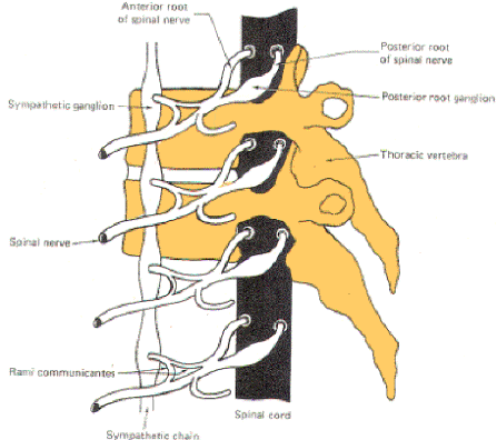
Fig-1: The sympathetic ganglia  associated with spinal nerves  T1 through L2 are connected to the nerve by two arms, the rami communicantes albicans and gresiumFig-2: There are no rami albicans above T1 and below L2. The gray rami send connections to all 31 nerves.Fig-3: The sympathetic outflow. The preganglionic neurons originate  in the intermediolateral horn of the spinal cord, between T1 and L2

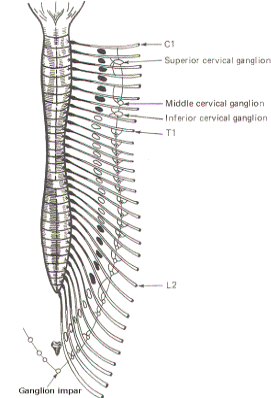
The sympathetic ganglia lie close to the vertebral bodies and are also known as paravertebral ganglia. They are strung together to form a sympathetic or paravertebral chain. There are two of these chains, one on either side of the vertebral column connected in front of the coccyx by the single ganglion impar (Fig-2).
Some of the fibers from nerve cells within the ganglia return to the spinal nerve trunk via a gray ramus. The fibers traveling through the white rami are myelinated while those in the gray rami are not, and this fact is responsible for their respective names. Each of the twelve thoracic and first two lumbar nerves is in contact with a paravertebral ganglion via a white and gray ramus. However, there are three ganglia in the chain above the thoracic region as well as several below L2 (Fig. 14-2). Each of these additional ganglia is connected to a spinal nerve by a single gray ramus.
The superior, middle, and inferior cervical ganglia probably represent the fusion of smaller individual cervical ganglia. These three send gray rami to all eight cervical spinal nerves. The superior cervical ganglion sends to the first four cervical nerves, the smaller middle cervical ganglion supplies the next two, and the large inferior cervical ganglion projects a gray ramus to the seventh and eighth cervical nerves. Similarly, a variable number of ganglia (four to eight) below L2 send gray rami to all of the spinal nerves below this level. Consequently, all 31 pairs of spinal nerves are in contact with the sympathetic chain and carry fibers of the sympathetic system. This is an important feature, enabling those effector organs which are innervated only by spinal nerves (cutaneous and skeletal muscle blood vessels, sweat glands, and pilomotor smooth muscle) to receive sympathetic input.
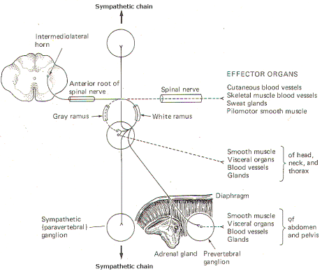
In addition to the paired paravertebral ganglia, there are several unpaired prevertebral ganglia in the abdomen and pelvis. They also playa role in the sympathetic outflow. Figure 14-3 illustrates the many possible ways by which the sympathetic system innervates its effector organs.
There is always a two-neuron link to each effector organ innervated. with the single exception of the adrenal medulla. The first is the preganglionic neuron and the second is the postganglionic neuron. The four possible routes of the preganglionic and postganglionic fibers, as illustrated in Fig. 14-3, are summarized below. After entering the sympathetic ganglia via the white rami, the preganglionic fibers may:
1 Pass without synapsing up or down the sympathetic chain to ultimately synapse in a higher or lower ganglion. By passing up the chain, the first four or five thoracic cord levels contribute all of the preganglionic fibers to the superior, middle, and inferior cervical ganglia. Similarly by passing down the chain, the lower thoracic and upper lumbar cord levels contribute all of the preganglionic fibers to the ganglia in the chain below L2. Postganglionic fibers then leave the ganglia via their gray rami to enter their respective spinal nerves for distribution to their effector organs (cutaneous and skeletal muscle blood vessels. sweat glands, and pilomotor smooth muscle).
2 Synapse in the ganglia and subsequently stimulate postganglionic fibers which leave the ganglia to reenter the spinal nerves via the gray rami. The postganglionic fibers are then distributed with the spinal nerves to their effector organs (cutaneous and skeletal muscle blood vessels. sweat glands, and pilomotor smooth muscle).
3 Synapse in the ganglia and subsequently stimulate postganglionic fibers which leave the ganglia and are directly distributed to their effector organs (smooth muscle, visceral organs. blood vessels, and glands of the head, neck, and thorax).
4 Pass without synapsing into the abdomen to synapse in one of the prevertebral ganglia or the adrenal medulla. Postganglionic fibers leave the prevertebral ganglia to innervate their effector organs (smooth muscle. visceral organs. blood vessels, and glands of the abdomen and pelvis).
THE PARASYMPATHETIC OUTFLOW

The nerve fibers which comprise the parasympathetic system originate in two quite distant regions, the brain stem and the sacral portion of the spinal cord. For this reason it is often called the craniosacral outflow to distinguish it from the thoracolumbar outflow of the sympathetic system. Those GVE fibers which make up the cranial portion of the system originate in specific brain stem nuclei and are distributed with cranial nerves III, VII, IX, and X. Those which comprise the sacral portion originate in lamina VII of sacral cord segments 2 to 4 and are distributed as the GVE fibers of the pelvic nerves (nervi erigentes). As with the sympathetic system, there are always two neurons in the pathway to the effector organ supplied. Thus, there are pre- and postganglionic fibers in the parasympathetic system also. However, unlike those in the sympathetic system, parasympathetic ganglia are quite distant from the brain stem and cord, often located directly on the effector organ itself. Thus the postganglionic fibers are much shorter in the parasympathetic system than they are in the sympathetic system. It should be noted here that autonomic effector organs typically receive both sympathetic and parasympathetic innervation, though some receive input from one system only. The effects of sympathetic and parasympathetic stimulation of the autonomic effector organs are summarized in Table-1. The effects are often but not always opposite, as will be described later.
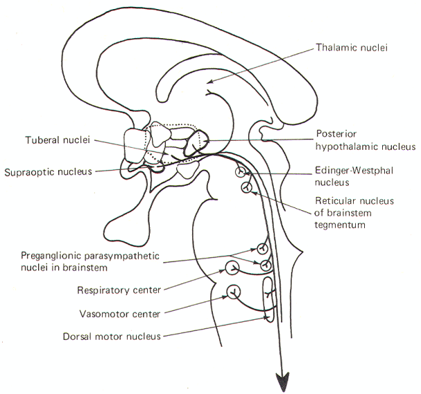
Figure-4 illustrates the parasympathetic outflow. The Edinger­Westphal nucleus (an accessory nucleus of III) in the tegmentum of the midbrain gives rise to the preganglionic parasympathetic fibers of the oculomotor (III) nerve. Some of these fibers terminate in the ciliary ganglion and others in the episcleral ganglion. The former stimulate postganglionic fibers innervating the sphincter muscles of the iris, which control pupillary diameter, while the latter stimulate postganglionic fibers innervating the ciliary muscle controlling the curvature of the lens (Fig-5).
The superior salivatory nucleus in the pons gives rise to the preganglionic fibers of the facial (VII) nerve. Some of these fibers terminate in the sphenopalatine ganglion and others in the submandibular (submaxillary) ganglion. Postganglionic fibers from the former innervate the lacrimal gland and mucous membranes in the head and neck region while postganglionic fibers from the latter innervate the submaxillary and sublingual salivary glands. The inferior salivatory nucleus at the pontomedullary border gives rise to the preganglionic fibers of the glossopharyngeal nerve (IX). These fibers terminate in the otic ganglion, from which postganglionic fibers innervate the parotid gland.
The overwhelming majority of cranial preganglionic fibers are distributed within the vagus (X) nerve. They originate in the dorsal motor nucleus of X in the medulla and terminate in unnamed peripheral ganglia on thoracic and abdominal organs, glands, and some blood vessels. Short postganglionic fibers run from these ganglia to receptor sites on the effector organ cells.
The sacral parasympathetic outflow supplies the organs and glands in part of the lower abdomen and all of the pelvis. Included are the descending colon, sigmoid, rectum, bladder, and external genitalia. As noted earlier, the preganglionic fibers originate in lamina VII of the sacral cord between S2 and S4, These fibers travel with the pelvic nerve and terminate in peripheral ganglia on the effector organs themselves.
Fig-4: The parasympathetic outflow
 

AUTONOMIC NEUROTRANSMITTERS

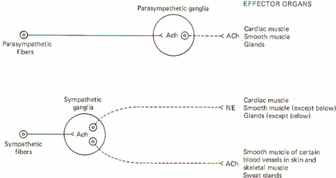
Both sympathetic and parasympathetic preganglionic neurons are cholinergic; that is, the preganglionic fibers of both systems release acetylcholine (ACh) at the synapse in the ganglion. Thus ACh is the principal transmitter in the autonomic ganglia. There are also some dopaminergic (dopamine releasing) interneurons present, but their function is still unknown. Nevertheless, the preganglionic fibers themselves are all cholinergic.
All postganglionic fibers of the parasympathetic system are cholinergic, but postganglionic sympathetic fibers are more diverse. The overwhelming majority areadrenergic [release norepinephrine (NE)], but a few are cholinergic. The few which are known to be cholinergic are those which innervate the sweat glands and some cutaneous and skeletal muscle blood vessels. (Fig-5)
Fig-5: General scheme of autonomic neurotransmitters.
 

Acetylcholine Synthesis, Release, and Inactivation
Figure-6 illustrates the general scheme of activity at the cholinergic synapse. Synthesis of ACh occurs in the cytoplasm of cholinergic presynaptic terminals. Coenzyme A (CoA) combines with acetate to form acetyl coenzyme A (acetyl CoA). Energy for this reaction is supplied by ATP. Once formed, the acetyl CoA combines with choline in the presence of the enzyme choline acetyltransferase to form acetylcholine (ACh). Once synthesized, ACh is taken up by the synaptic vesicles and held there in a bound form until its released.
When an impulse reaches the presynaptic terminal, several synaptic vesicles release ACh into the synaptic cleft. ACh then diffuses across the cleft to activate cholinergic receptor sites on the postsynaptic membrane. In order to allow the presynaptic terminal to effectively control the postsynaptic membrane, the released ACh must be quickly degraded (within microseconds) by the enzyme acetylcholinesterase (AChE) to acetate and choline, which are then reabsorbed into the presynaptic terminal for resynthesis to ACh. A small fraction is reabsorbed intact into the presynaptic terminal while an even smaller fraction diffuses out of the synaptic cleft before it can be degraded or reabsorbed. AChE is abundantly available in the cholinergic synaptic cleft. And even though the enzyme can degrade ACh within microseconds, there is adequate time for the ACh to activate receptor sites.Norepinephrine Synthesis, Release, and Inactivation.
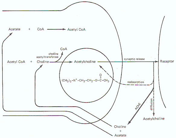
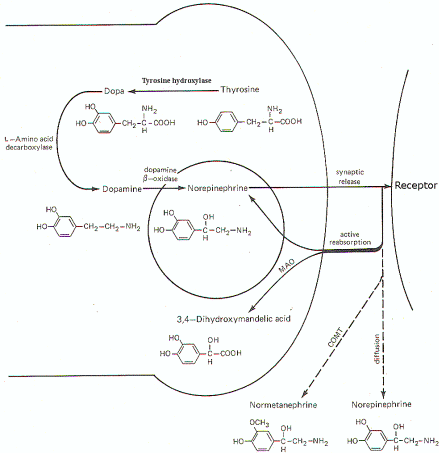
Fig-6: Synthesis and fate of synaptically released acetylcholine at cholinergic synapse.Fig-7: Synthesis and fate of synaptically released norepinephrine at adrenergic synapse.
Figure-7 illustrates the synthesis and fate of synaptically released norepinephrine at adrenergic synapses. Norepinephrine is synthesized in the presynaptic terminal by a series of enzymatically catalyzed reactions typically starting with the amino acid tyrosine. The sequence can also start with phenylalanine, which can be enzymatically converted to tyrosine. In either case tyrosine is converted to dihydroxyphenylalanine (dopa), dopamine, and finally to norepinephrine. The final synthetic step from dopamine to norepinephrine occurs in the synaptic vesicle where the norepinephrine is held in a bound form. The formation of dopa is apparently the rate-limiting step in the synthesis of norepinephrine. When an impulse reaches the presynaptic terminal, several vesicles release norepinephrine into the synaptic cleft, where it diffuses to activate receptor sites on the postsynaptic membrane. Within a few milliseconds, the norepinephrine is subject to one of three fates. A small amount is methylated by the enzyme catechol-o-methyl transferase (COMT), which is present in the cleft, and thereby rendered inactive. An even smaller fraction diffuses out of the cleft and away from receptor sites. But certainly the greatest amount of norepinephrine is reabsorbed by active transport into the presynaptic terminal. If norepinephrine stores in the synaptic vesicles are low, as might be the case in a rapidly firing fiber, the reabsorbed norepinephrine may be taken up by the vesicles for subsequent rerelease. If adequate stores of the transmitter are available, the reabsorbed norepinephrine is subjected to oxidative deamination by mitochondrial monoamine oxidase (MAO).
AUTONOMIC TONE
Table1 shows the effects of sympathetic and parasympathetic stimulation on autonomic effector organs. The sympathetic and parasympathetic systems are continually active and the level of activity at a given rate of firing is known as autonomic tone.

Table-1 Autonomic Effects on Various Organs of the Body
Effector organsEffects of sympathetic stimulationEffects of parasympathetic stimulation
EyeRadial muscle of the iris(α) Contraction (mydriasis) 
 Sphincter muscle of the iris Contraction (myosis)
 Ciliary muscle of the lens(β) Relaxation Lens flattensContraction ( Lens curves)
HeartSA node(β) ↑ heart rate↓ heart rate
 Atria(β) ↑ heart rate and force↓ heart force
 AV node(β) ↑ conduction velocity↓ conduction velocity
 Purkinje system(β) ↑ conduction velocity 
 Ventricles(β) ↑ heart rate and force 
Blood vesselsCoronary(α) ConstrictionDilatation
  (β) Dilatation 
 Cutaneous(α) Constriction 
  (ACh) Dilatation 
 Skeletal muscle(α) Constriction 
  (β) Dilatation 
  (ACh) Dilatation 
 Abdominal visceral(α) Constriction 
  (β) Dilatation 
 Renal(α) Constriction 
 Salivary glands(α) ConstrictionDilatation
StomachMotility and tone(β) Decrease (usually)Increase
 Sphincters(α) Contraction (usually)Relaxation (usually)
 SecretionInhibition (?)Stimulation
IntestineMotility and tone(α, β) DecreaseIncrease
 Sphincters(α) Contraction (usually)Relaxation (usually)
 SecretionInhibition (?)Stimulation
Gallbladder and ducts RelaxationContraction
Urinary bladderDetrusor(β) Relaxation (usually)Contraction
 Trigone and sphincter(α) ContractionRelaxation
UreterMotility and toneIncrease (usually)Increase (?)
Male sex organs EjaculationErection
SkinPilomotor muscles(α) Contraction 
 Sweat glands(α) Slight, localized secretions 
  (ACh) Generalized secretions 
Spleen capsule (α) Contraction 
Lung (bronchial muscles) (β) RelaxationContraction
Adrenal medulla  Secretion of epinephrine and norepinephrine
Liver (β) Glycogenolysis 
PancreasAcinar cells↓ secretionSecretion
 Islet cells(α) Inhibition of insulin and glucagon secretionInsulin and glucagon secretion
  (β) Insulin and glucagon secretion 
Salivary glands (α)  Thick, sparse secretionProfuse, watery secretion
Lacrimal glands  Secretion
Nasopharyngeal glands  Secretion
Adipose tissue (β) Lipolysis 
Juxtaglomerular cells (β) Renin secretion 
Pineal gland (β) Melatonin synthesis and secretion 

Sympathetic Tone
To illustrate sympathetic tone, consider this example. Most arteries are normally in a state of partial constriction. That is, they are neither fully constricted nor fully dilated. Since most blood vessels receive only sympathetic innervation, it is the only system that need be considered. If the normal partially constricted state of an artery is maintained by a basal firing rate of 1 impulse per second, we can describe the artery as displaying a basal sympathetic tone. Now if the firing rate should increase to say 50 impulses per second, the artery would constrict further, showing an increase in sympathetic tone. Conversely, if the firing rate were to decrease, the smooth muscle of the blood vessel would relax, causing the artery to vasodilate with a decrease in sympathetic tone.
The adrenal medulla is also an important contributor to sympathetic tone throughout the body. Each time the sympathetic system is activated, the adrenal medullae are also sufficiently stimulated via the splanchnic nerves, to increase their output of epinephrine and norepinephrine to the general circulation. These two catecholamines then travel to all parts of the body stimulating sympathetic effector organs. It is easy to see how the increased release of these two chemicals by the adrenal medulla can cause a general increase in sympathetic tone throughout the body. In fact. this increased output by the adrenal gland with sympathetic stimulation is the principal reason why the effects of sympathetic stimulation are longer lasting and more diffuse than those associated with the parasympathetic system.
Parasympathetic Tone

An example of parasympathetic tone is the control of peristalsis in the GI tract. Gastrointestinal smooth muscle receives both sympathetic and parasympathetic innervation. Increasing the firing rate of parasympathetic fibers to the gut causes an increase in intestinal motility and peristalsis, and hence, an increase in parasympathetic tone. Decreasing the firing rate produces a decrease in peristaltic activity, and hence, parasympathetic tone. Table-1 shows parasympathetic stimulation increases peristalsis while sympathetic stimulation decreases it. Thus, the GI musculature is an example of the often true observation that the effects of sympathetic and parasympathetic stimulation are opposite and tend to balance each other. Further examination of Table-1 , however, will show that this is not always true.
Alpha and Beta Receptors
The action of catecholamines on adrenergic effector organs varies with the organs. Catecholamines excite some effectors and inhibit others. Experiments with a series of sympathetic drugs have shown there are at least two types of adrenergic receptors. They are called alpha and beta. Blocking agents were later developed for each receptor which further confirmed their existence. The response of an effector to a catecholamine is then partly a function of the type of receptor it has. Epinephrine excites both alpha and beta receptors quite equally, while norepinephrine excites mainly alpha receptors. Nevertheless, norepinephrine will also excite beta receptors, but only to a slight extent. This explains why epinephrine has a much stronger effect on the heart (which has only beta receptors) than norepinephrine does. To further confuse the picture, some effectors have only alpha receptors, others have only beta receptors, and still others have both. Thus the specific response of an effector is both a function of the relative ratio of receptor types and the kind of transmitter involved. A partial list of the effects of alpha and beta stimulation is given in Table-2.
Table-2 Effects of Alpha and Beta Stimulation
Alpha receptorBeta receptor
VasoconstrictionVasodilation
Mydriasis (pupil dilation)Cardioacceleration
Intestinal relaxationBronchial relaxation
 Increased cardiac strength
 
Glycogenolysis
 Lipolysis
Notice that some alpha functions are inhibitory while others are excitatory. The same is true for certain beta effects. Therefore it is not possible to refer to one receptor as excitatory and the other as inhibitory, as is sometimes true. Beta receptors have also been divided into two types: beta1 and beta2, according to their responses to various drugs. Beta1 receptors are those responsible for the inotropic (strength) and chronotopic (rate) responses of the heart. as well as lipolysis. Beta2 receptors bring about vasodilation and bronchial relaxation. This is a distinction useful to the pharmacologist, who can then use a beta2 agonist to treat asthma and produce bronchial relaxation with very little cardiac stimulation.
AUTONOMIC AND RELATED DRUGS
A large number of drugs have been developed which are active at various sites in the autonomic nervous system. Figure 10 schematically illustrates the action and site of action of several of these.
 
Fig-8: Action of various drugs on the autonomic nervous system.
Drugs Acting on Autonomic Effector Organs
Acetylcholine, pilocarpine, and methacholine all directly stimulate cholinergic receptors on autonomic effector organs. Physostigmine and neostigmine also potentiate activity at these receptors, but do it by the indirect action of inhibiting cholinesterase (AChE). Conversely, atropine is a potent antagonist at these receptors, inhibiting the action of endogenously released ACh as well as administered cholinomimetic drugs.
A variety of drugs are also active at adrenergic receptors on autonomic effector organs. Norepinephrine, epinephrine, isoproterenol (a beta agonist) and phenylephrine (an alpha agonist) are all capable of directly stimulating these receptors. In addition, ephedrine and metaraminol can act directly on these receptors but typically are first absorbed by the adrenergic nerve endings and subsequently released upon the arrival of impulses at the presynaptic terminal. Metaraminol is an alpha agonist both directly and indirectly, while ephedrine is a beta agonist directly but stimulates alpha receptors when released by adrenergic nerve endings. On the other hand, phentolamine and phenoxybenzamine are effective alpha antagonists and thus effectively block alpha receptors. Propranolol is a beta blocker.
Drugs Acting on Autonomic Nerve Endings

There are no known drugs to stimulate the release of ACh from the presynaptic terminals of cholinergic nerve endings. However, botulinum toxin is a potent inhibitor of ACh release. Adrenergic nerve endings are more commonly manipulated by drug action. Both tyramine and amphetamine promote the release of endogenous norepinephrine from these nerve endings. Ephedrine and metaraminol are also potentiators at these sites by the indirect action of being absorbed into the terminals and subsequently being released as false transmitters. Reserpine and guanethidine are effective inhibitors here by the action of depleting stores of norepinephrine in synaptic vesicles and preventing their further uptake and storage.
Drugs Acting on Autonomic Ganglia

Drugs active at parasympathetic ganglia are equally effective at sympathetic ganglia, and vice versa. Nicotine stimulates postganglionic neuron receptors in the autonomic ganglia. Hexamethonium and mecamylamine effectively block these "nicotinic" receptor sites.
Nonautonomic Drugs

It is worth pointing out that there are several drugs which are active at the skeletal neuromuscular junction which are not active in the autonomic nervous system. For example, curare and succinylcholine effectively block the action of ACh on skeletal muscle receptors but have no similar ACh blocking action on cardiac, and smooth muscle receptors.